


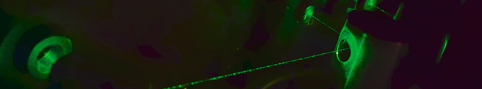


Wide field of view image of the tubulin cytoskeleton of U2OS cells acquired by waveguide-based dSTORM. This images was obtained by using a waeguide chip for TIR illumination and an air objective lens to collect the fluorescence. Several throusands of images were collected and then analyzed using "ThunderSTORM".
Wide field-of-view imaging by separated illumination and detection
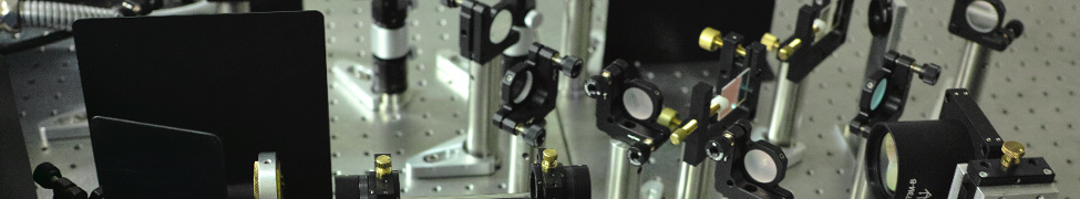
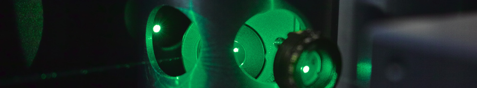
In order to achieve optimal sample illumination, single-molecule localization microscopy techniques typically rely on illuminating the sample using oil objective lenses with high numerical aperture that also serve as the fluorescence detection optics. These setups consequently yield small fields of view and are not very flexible with regard to the choice of objective lenses. In our research we have pioneered the use of planar optical waveguides as sample holders that also provide sample illumination for super-resolution optical microscopy. A laser light source is coupled into a high-refractive index optical waveguide (Ta2O5or SiN3, or other high index transparent materials) either from the side on the end-facet, or by using a grating coupler etched into the chip. The light then progresses through the waveguide by total internal reflection (TIR), and the induced evanescent field excites the fluorophores in the sample close to the waveguide-sample boundary.
Using this technique we are able to decouple illumination from the detection pathway, giving us the liberty to choose any objective lens we want, and still achieve TIR-illumination. We have shown that it is possible to combine waveguide-illumination with super-resolution optical imaging techniques such as direct Stochastic Reconstruction Microscopy (dSTORM), using low numerical aperture objective lenses. The result yields cell structures at nanometer resolution with a field of view of up to half of a millimeter with excellent signal to noise ratio.
By combining waveguide-illumination with our research on cost-effective microscopy setups using industry-grade complementary oxide-semiconductor cameras, we hope to potentially turn any microscope into a optical nanoscope.

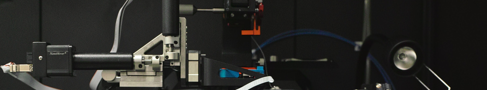
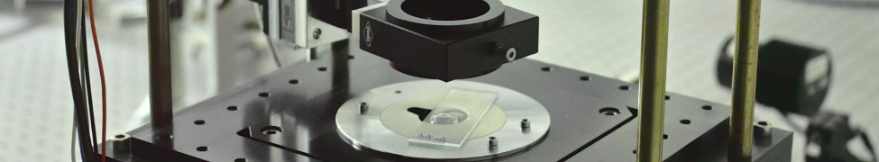
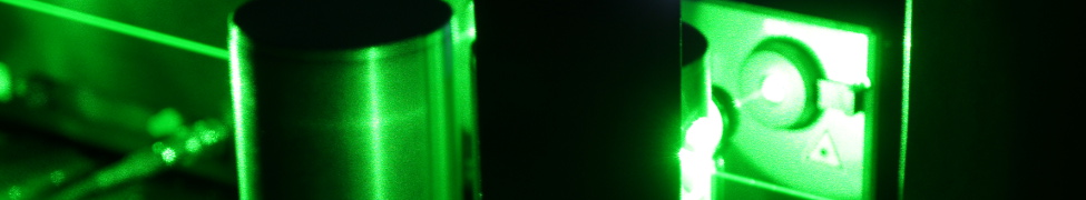
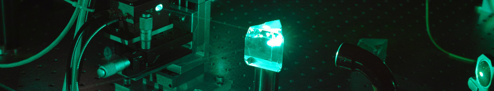
Optical waveguide chip with 647 nm light coupled in from the side. Above the chip an objective revolver can be seen with several air objective lenses which serves as detection optics.
Publications/Further reading:
Chip-based wide field-of-view nanoscopy
Diekmann R, Helle OI, Oie CI, McCourt P, Huser T, Schüttpelz M, Ahluwalia BS
Nature Photonics 11(5): 322-328 (2017)
PUB| DOI| WoS
Characterization of an industry-grade CMOS camera well suited for single molecule localization microscopy – high performance super-resolution at low cost
Diekmann R, Till K, Müller M, Simonis M, Schüttpelz M, Huser T
Scientific Reports 7(1): 14425 (2017)
PUB| PDF| DOI|WoS| PubMed| Europe PMC