


Implementing fluorescence microscopy and super-resolution optical microscopy in cost-efficient implementations
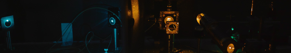
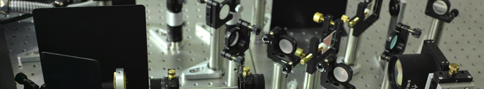
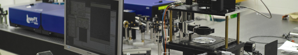
In our research group, we are developing and operating several compact fluorescence microscopy systems. One of the main advantages of these systems is their easy transportation to remote sites or to the laboratory of collaborators in biology, medicine, or biochemistry. It is often easier to ship a powerful optical setup, rather than to transport delicate living biological samples. Additionally, a compact design urges the microscope builder to uses small cameras. In our case, we are using industry grade CMOS cameras which are also very cost-effective, which makes building compact microscopy systems even more attractive. These industry grade cameras can compete with scientific grade cameras and deliver comparable results. For now, we just describe a few of these systems, but we plan to also produce and release open access blueprints for such systems, once they have proven to be robust and easy to reproduce, to allow other researchers to build their own systems.
Every biological or medical application of high resolution optical microscopy creates new and different challenges. In one case, the optical resolution might be important, while in another case the acquisition speed is of more interest. Therefore, we developed different compact fluorescent microscopy systems to be able to investigate a wide variation of important medical and biological samples: Our compact deconvolution widefield fluorescence microscopy setup (cDecon) enables the observation of fast dynamics in living cells in 3D. If more resolution is required to investigate fixed sample, then our compact dSTORM setup (cSTORM) is the way to go. If, however, a problem requires super-resolution optical imaging and high-speed image acquisition, then Structured Illumination Microscopy (SIM) is the most suitable method. Right now, a suitable setup for compact SIM (cSIM) is under construction.
cDecon
A point-emitter in a fluorescence microscope appears as a so-called Airy disk which can be described by the Point Spread Function (PSF). Mathematically speaking, the sample is convoluted with the PSF of the microscope, resulting in diffraction-limited images. In widefield fluorescence microscopy, this causes background blur and further reduces the optical resolution. To overcome these issues the image can be transformed to Fourier space and then deconvolved with the Optical Transfer Function (OTF) of the microscope system, which is the PSF in Fourier space.
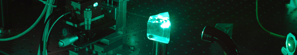
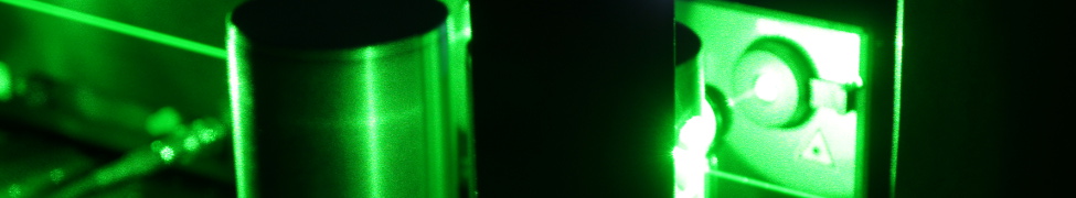
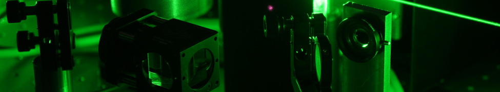
For applying deconvolution, a normal widefield fluorescence microscope is suitable. In our case, we additionally removed superfluous components to achieve a compact design. The current system is stable enough to remove the need for an expensive air-compensation table. Placing the system on top of the inner tube of a bicycle tire is sufficient to create sufficient vibration isolation. With this system, two fluorescent probes can be observed and tracked simultaneously, e.g. GFP and mCherry. These are fluorescent proteins which are commonly used in biological and medical applications.

The first iteration of the setup was shipped to the laboratory of our collaborator, Prof. Benjamin Chen at the Icahn School of Medicine at Mount Sinai in New York City, NY, to image the transfer of HIV in a biosafety lab. Jurkat T cells were transfected with plasmids encoding HIV proteins which were fused to GFP and mCherry. We imaged living cells for extended periods of time and were able to observe virus transfer between cells.
Further projects, such as imaging living cardiomyocytes (heart muscle cells), performing live deconvolution reconstruction, etc. are currently in progress.
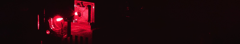
cSTORM
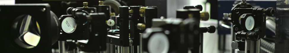
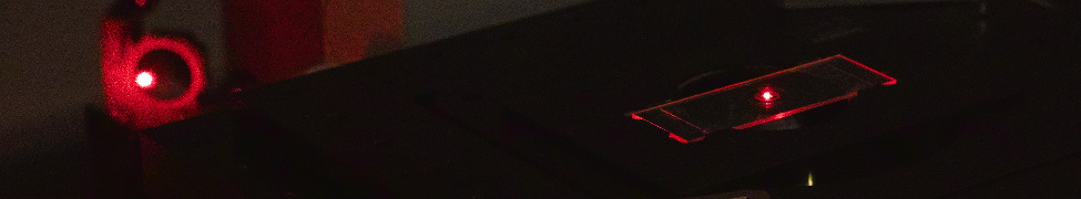
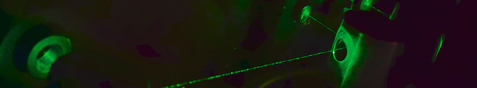
This compact and cost-efficient dSTORM set-up fits on a breadboard of 44x36cm. Illumination methods, such as HILO and TIRF are achieved by lateral beam displacement via a translation stage. The set-up is easy-to-use and inexpensive, and can therefore also be used to explain and demonstrate the handling and understanding of SMLM techniques, such as dSTORM, e.g. in local high schools or during public outreach events. This system is frequently used to explain the principles of single-molecule localization microscopy and to explain the basic microscope optics.

Due to the compactness and robustness and the use of a small, uncooled industry grade CMOS camera (IDS μeye) with an USB 3 connection this system is highly mobile and very robust. This was proven by shipping it to another laboratory in Spain and performing measurements there as part of our participation in the Tollerrant Innovative Training Network. Moreover, alterations and extensions can easily be made. A similar version with two laser lines was set up in a laboratory of our collaborators at the Centre for Education and Research in Ageing in Sydney, Australia.
In addition to these attributes, the cSTORM system also displays high performance, which was shown by a direct experimental comparison between the industry-grade CMOS camera and a commonly used scientific CMOS camera (Hamamatsu Orca Flash) 1. Even though minute differences in the technical performance of the industry grade CMOS camera were found when comparing it to an sCMOS camera, similar spatial resolution was obtained in superresolved microscopy images of real samples (shown beneath) for both cameras. 1

Right: Reconstructed dSTORM images of Alexa 647 immunostained microtubules in fixed U2OS cells.1
Left: The spatial resolution as obtained by Fourier Ring Correlation analysis for both cameras is on the order of 38nm. 1
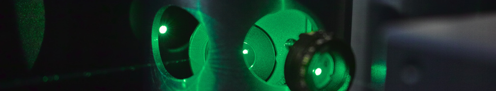
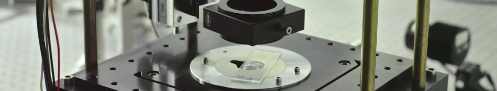
cSIM
The basic principle of SIM is the illumination of the sample with a sinusoidal excitation light pattern, ideally an interference pattern with a periodicity near the optical diffraction limit. Therefore, unresolved spatial frequencies can be shifted to resolvable spatial frequencies, which can be imaged by the microscope. Using computational image reconstruction2the original image can be calculated and reconstructed to a super-resolved image.
For the realization in real-life applications there exist different approaches to generate the sinusoidal intensity pattern. In our lab, we are utilizing two-beam interference pattern which are generated by Spatial Light Modulators (SLM). The SLM displays typically optical gratings which result in an interference pattern when the different beams diffracted by the grating pattern are projected into the sample by suitable lenses. For the calculation of the reconstructed image the pattern, typically at least 9 raw images (three angles and three different phases per angle) are required for 2D image reconstruction and 15 raw frames (of a 3D interference pattern) are required for 3D image reconstruction. Therefore, the SLMs are typically electrooptical devices that can rapidly generate the different grating patterns customized for each excitation wavelength.
In our laboratory, we are currently designing a compact SIM which can also easily be transported to biosafety laboratories. Therefore, we have reduced the size of the overall system, used cost-efficient components (employing e.g. the same industry grade cameras as above) and simplified the overall operation of the system. A custom-written GPU-accelerated code for SIM image reconstruction is suitable for the real-time operation of this kind of system, and overcomes drawbacks compared to the high cost and complicated operation of conventional SIM systems.

References:
(1) R. Diekmann, K. Till, M. Müller, M. Simonis, M. Schüttpelz, and T. Huser, Characterization of an industry-grade CMOS camera well suited for single molecule localization microscopy – high performance super-resolution at low cost, Sci. Rep.7, 14425 (2017)
(2) M. Müller, V. Mönkemöller, S. Hennig, W. Hübner, and T. Huser,Open source image reconstruction of super-resolution structured illumination microscopy data in ImageJ, Nature Commun. 7, 10980 (2016), DOI: 10.1038/ncomms10980